Typical flow cytometer consists of five basic operational units:
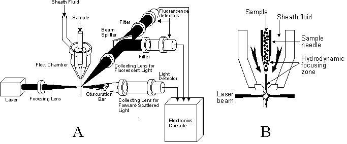
Schematic view of a flow cytometer (A) and a close up of the flow chamber (B) (adapted after Doležel 1997)
Two types of light sources are used in plant flow cytometers: lasers and/or arc lamps.
Lasers produce stable, bright, narrow beam of monochromatic light. Its wavelength depends on a filled gas in the plasma tube. Majority of current cytometers work with air-cooled argon ion laser tuned at 488 nm (turquoise), although other types are also commercially available (e.g. diode laser, solid state laser, etc.). Spectral purity of the emitted light eliminates the need for excitation filters, however, it simultaneously represents one of the major limitations. If other wavelengths are required, another laser should be used. High price is additional laser disadvantage.
Mercury (or rarely xenon) arc lamps are employed as an inexpensive source of light in some bench-top instruments. In plant sciences, they are well suited particularly for ploidy level estimation. Low cost and easy maintenance rank among their principal advantages over lasers. They emit non-monochromatic light, therefore a set of excitation optical filters must be used to select required wavelength (usually in UV). Mercury lamps also suffer from rather low lifetime (generally between 200 - 400 hours) and a low output power (insufficient sensitivity to analyse a weak fluorescence). Moreover, gradual decrease in light intensity within a working day can occur.
Flow chamber (= flow cell, nozzle) represents central part of the instrument. Its mission is to adjust the measured particles in a narrow central stream and to deliver them one after another into a focal point of the light source. This is achieved by so-called hydrodynamic focusing: the sample is injected into a stream of sheath fluid (mostly water or saline solution) moving with a greater velocity and thus confining the sample within a central core. Induced acceleration at the narrowing flow chamber orifice forces the particles to move singly, and they are delivered to the point of excitation. Typical stream velocity is between 1 and 10 m/s that corresponds to several dozens or hundreds of analysed particles per second. There are three basic types of flow chamber configuration. In 'jet-in-air' design, the stream exits from the flow chamber into open air where the particles intersect a light beam. As the flow chamber orifice has a small diameter (typically 75 µm), high speed is attained. 'Enclosed stream' design operates with lower velocity (higher sensitivity is often achieved), and the particles are measured during their movement in a narrow capillary tube (of about 250 µm in cross-section). In 'jet on open surface' design, the particles are analysed in a stream flowing on a glass coverslip
Optical part of the instrument provides focusing the excitation beam, selection of required wavelengths, collection of output light and its delivery to the detectors. Non-adjusted laser beam has a circular shape of approximately 1-2 mm in diameter. However, to achieve identical illumination of each particle, it must be focused with lenses to an appropriate profile. Elliptical spot of about 60 × 20 µm (with longer dimension perpendicular to the sample stream) is preferred for DNA amount analysis in 'enclosed stream' cytometers. The output light from illuminating particles (collected through the lens with a high numerical aperture) consists of various colours. This spectral mixture must be partitioned into specified wavelengths before reaching the photodetectors. An assembly of optical mirrors, dichroic mirrors, and colour filters is used in this mission. Standard filters accommodated in flow cytometers are the short-pass ones (transmit light below a specified wavelength), longpass filters (transmit light above a specified wavelength), and band-pass filters (transmit light over a narrow wavelength band close to the specified value; they are generally used immediately in the front of the detectors). Dichroic mirrors (beam splitters) are placed at a 45° angle in the light path and they work in a similar fashion as filters (reflect vs. transmit light of specified wavelengths).
The light beam adjusted by the optical bench is focused onto photodetectors that convert light signal into electrical current impulse. After preamplification and further processing (e.g. elimination of debris and electric noise), the signal undergoes a main amplification. Both linear and logarithmic amplifiers are available in commercial flow cytometers; the former should always be used for nuclear DNA content estimation. Final stage before storing the signals, is an analogue to digital conversion. Ten bits converters yielding 1 024 channels are most commonly used in DNA measurements.
Digital data are stored, visualised, and further analysed in a built-in computer. Majority of systems use a flow cytometry standard (FCS) format for the data storage. On the computer screen, they are generally displayed as univariate or bivariate histograms. Often, it is necessary to select certain subgroup of particles before performing statistic analyses (procedure called gating). Once stored, the data can be subjected to various statistic procedures to extract required information. Mean channel position, number of particles, and coefficient of variation are the basic statistics computed in univariate analyses.